


- Home
- Hair medicine
- Hair loss
- Trichoscopy
Trichoscopy: the examination that shows the scalp
What trichoscopy is, what it shows in each type of hair loss, how the examination is done and why it helps to monitor treatment.
 Rúben BritoAuthor
 Dr Michelle CampagnoliClinical review · OM no. 69467
In this article
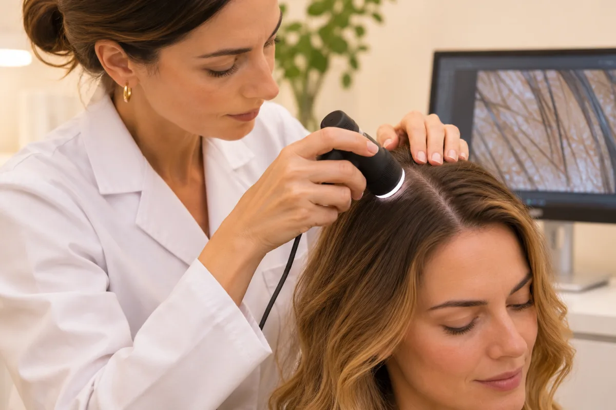
Trichoscopy is an examination that observes the scalp and hair shafts with a high-magnification lens. It shows details that cannot be seen with the naked eye: the thickness of each hair, signs of inflammation, empty follicles or hairs just emerging. It is quick, painless and needs no special preparation.
It serves two purposes: helping to understand the cause of hair loss and monitoring, with comparable images, whether treatment is working. The European guideline for androgenetic alopecia recommends it in cases of doubt (Kanti et al., 2017).
Three ideas to remember.
Fica por baixo do resumo, se existir uma segunda foto.
What it is and how it works
The device used is a dermatoscope, the same type of instrument used to examine moles on the skin, or a videodermatoscope connected to a computer. It magnifies the image between 10 and 70 times, or more, and uses its own light to eliminate glare.
With this magnification it is possible to assess:
- the hair shafts: thickness, differences in calibre between hairs, broken hairs or hairs with an abnormal shape;
- the follicle openings: whether they are present, empty or gone;
- the scalp skin: redness, scaling, vessels, pigmentation around the hairs;
- the follicular units: how many hairs come out of each pore. On a healthy scalp, many pores have 2 to 3 hairs.
It is different from the trichogram, an older test in which about 50 hairs are plucked to be observed under a microscope. Trichoscopy has replaced it in most situations because it is non-invasive.
What trichoscopy shows in each type of hair loss
| Condition | Typical signs on trichoscopy |
|---|---|
| Androgenetic alopecia | Hairs of very different calibres from one another; a variation of 20% or more is considered diagnostic. More pores with a single hair at the front than at the nape. Thin, short hairs (vellus). Yellow dots in advanced cases |
| Telogen effluvium | Short, straight new hairs scattered across the whole scalp, with no difference between the front and the nape. Trichoscopy serves mainly to exclude other causes |
| Alopecia areata | Black dots (hairs broken at the surface) and "exclamation mark" hairs, thinner at the base, which indicate active disease; yellow dots |
| Trichotillomania (hair pulling) | Hairs broken at different lengths, in the same area |
| Scarring alopecias | Absence of follicle openings, redness or scaling around the hairs. These need prompt assessment |
Source: DermNet; Rakowska et al., 2009.
The example of female hair loss
In women, telling androgenetic alopecia from chronic telogen effluvium is difficult with the naked eye, because both cause diffuse thinning. A study of 131 women proposed trichoscopic criteria that compared the front area with the nape: thinner hairs at the front, more than 10% of hairs under 0.03 mm and yellow dots. With these criteria, specificity for female androgenetic alopecia was 98% (Rakowska et al., 2009).
Trichoscopy does not replace the clinical history and, at times, blood tests or a biopsy. But it greatly reduces uncertainty.
How the examination is done
- Initial conversationThe doctor asks when the hair loss began, what changed in the previous months, medicines and family history.
- General observationof the scalp and, often, a gentle pull test.
- TrichoscopyThe device is placed against the scalp in several areas, usually the front, the top, the crown and the nape, which serves as a comparison. It does not hurt and no hair needs to be cut.
- Image recordingto compare at later appointments.
In total, the examination itself usually takes 10 to 20 minutes.
How to prepare
- Wash your hair as usual, preferably 1 to 2 days beforehand. A freshly washed scalp can hide scaling; too long without washing makes observation harder.
- Do not apply styling products, oils, hair fibres or scalp make-up on the day.
- Avoid colouring your hair in the previous week, if possible.
- Bring the list of medicines and supplements and, if you have them, recent blood tests and old photographs of your hair.
Monitoring progress
One of the biggest advantages of trichoscopy is that it allows comparison. Treatments for hair loss take months to show results, and what you see in the mirror can be misleading. Repeating the images in the same areas, at the same magnification, makes it possible to see:
- whether the hairs are getting thicker;
- whether there are more hairs per pore;
- whether new hairs are appearing;
- whether inflammation has improved.
As a rule, reassessment is done after 3 to 6 months of treatment. At home, you can complement it with regular photographs; we explain how in Photographs every 3 months: how to monitor the crown.
Limits of the examination
- The result depends on the experience of whoever interprets it.
- In telogen effluvium, the signs are not very specific: trichoscopy mainly helps to exclude other causes.
- In some scarring alopecias a biopsy is needed to confirm the diagnosis.
- It does not replace blood tests when deficiencies or hormonal changes are suspected. See Blood tests for hair loss: what to ask for.
When trichoscopy makes sense
It also makes sense before starting treatment, to have a starting point, and during treatment, to confirm that it is working.
At Luxmed, trichoscopy is part of the Hair Medicine consultation and is the basis for diagnosis and follow-up.
Talk to us
The assessment is a conversation, with no obligation. Tell us what concerns you and our team will reply.
Telephone 214 044 241 · info@luxmed.pt
This article is for information only and does not replace a medical consultation.
Frequently asked questions
Does trichoscopy hurt?
No. The device only touches the scalp. No hairs are plucked or cut.
How long does it take?
The observation itself takes 10 to 20 minutes, as part of an appointment.
Do I need to go with clean hair?
Ideally wash it 1 to 2 days beforehand and do not use styling products on the day of the examination.
Does trichoscopy replace blood tests?
No. It shows what is happening on the scalp; blood tests show deficiencies or hormonal changes. They are often complementary.
How often is it repeated?
As a rule, after 3 to 6 months of treatment and then once or twice a year, depending on progress.
Is a "salon" hair analysis the same thing?
Not necessarily. What sets medical trichoscopy apart is the clinical interpretation of the signs, combined with the history and tests, to reach a diagnosis.
Scientific references 4
- Rakowska A et al. Dermoscopy in female androgenic alopecia: method standardization and diagnostic criteria. Int J Trichology. 2009;1(2):123-130. Artigo
- DermNet. Trichoscopy of generalised noncicatricial hair loss. Artigo
- Kanti V et al. Evidence-based (S3) guideline for the treatment of androgenetic alopecia in women and in men. European Dermatology Forum, 2017. Artigo
- Trichoscopy of androgenetic alopecia: a systematic review. J Clin Med. 2024;13(7):1962. PubMed
Keep exploring