


- Home
- Hair medicine
- Thinning hair at the crown
- Photographs of the crown
Photos every 3 months: how to track the crown
Four photographs, always the same, every three months: how to build at home an album that shows real progress and that is useful at the consultation.
 Rúben BritoAuthor
 Dr Michelle CampagnoliClinical review · OM no. 69467
In this article
Anyone worried about their crown, or who has started a treatment for hair loss, wants to know one thing: is it getting worse, staying the same or improving? The mirror does not answer that question well. Photographs do, if they are always taken in the same way and at the right interval.
In clinical trials of treatments for hair loss, standardised photography is one of the main instruments for measuring results. At home, with a mobile phone and a few rules, it is possible to build an album that shows real progress and that is very useful at the consultation.
Three ideas to remember.
Fica por baixo do resumo, se existir uma segunda foto.
Why every three months
The rhythm of hair
Hair grows about 1 centimetre a month, and each follicle goes through growth and resting phases that last months. The resting phase lasts about three months. That is why any change in density, for better or worse, only becomes visible after several months.
The pace of treatments
Treatments with evidence for androgenetic alopecia, such as minoxidil and finasteride, take months to show an effect. The European guideline (Kanti et al., 2018) recommends assessing the response after about six months and, preferably, twelve. Before that, photographs can mislead.
The right balance
- More often than every three months: it shows no differences and feeds anxiety.
- Less often than every six months: you miss the start of a change.
- Three months: four points a year, enough to see a trend.
A good idea is to link the date to something fixed: the start of each season, or the week after a haircut.
The protocol
Material
- Phone with a rear camera.
- Someone to help, or a tripod and the timer.
- A chair, always the same one, next to a window with diffuse natural light.
- A fine comb.
- A mark on the floor (adhesive tape) for the position of the chair.
Preparation
- Hair washed the day before and dry, with no gel, hairspray, wax, fibres or coloured spray.
- Approximately the same length: for example, always one to two weeks after a haircut.
- Same colour: if you colour your hair, photograph at the same point in the colouring cycle; freshly coloured hair disguises more.
- Style your hair as usual, without pulling.
The four photographs
| No. | View | How |
|---|---|---|
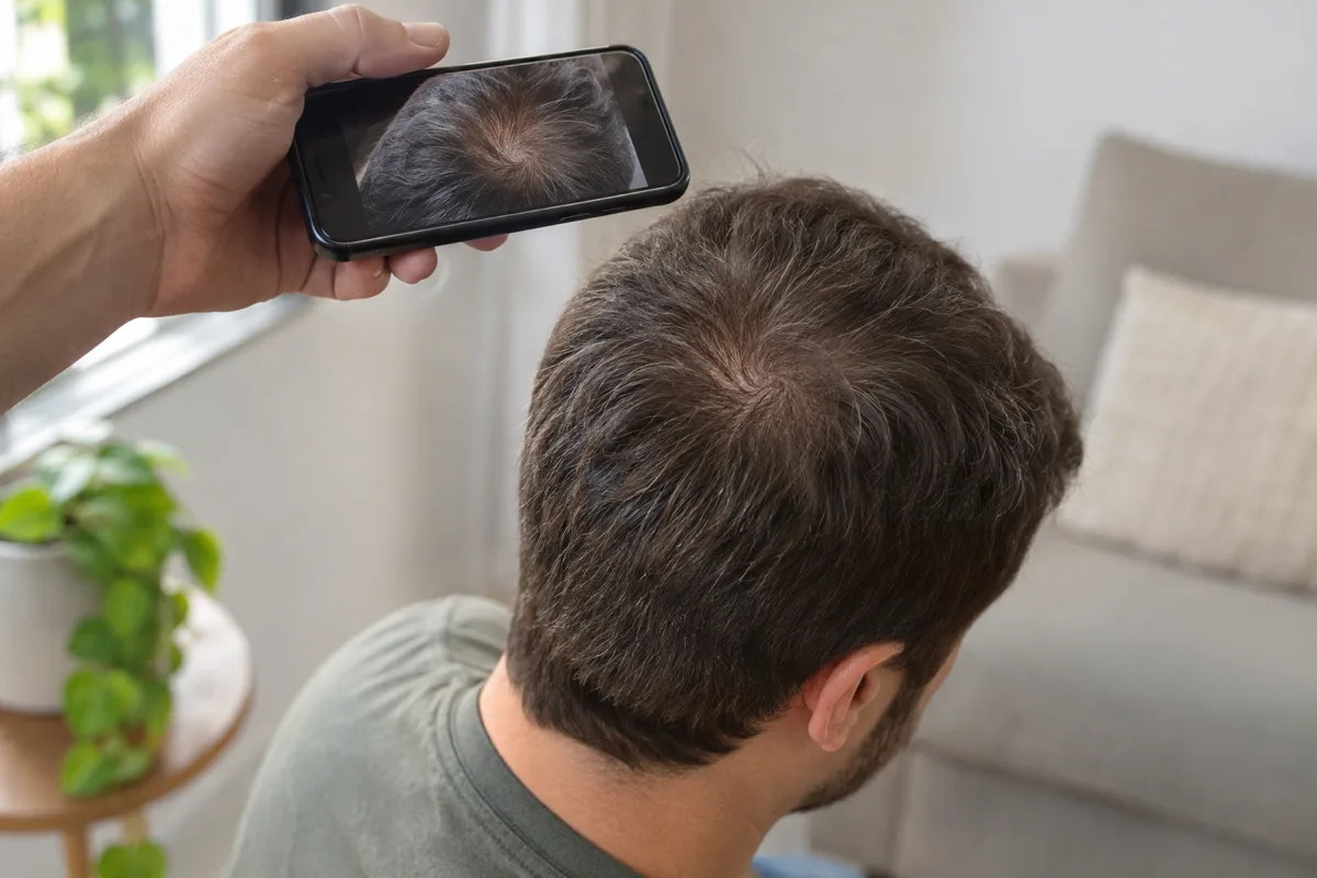
| 1 | Crown | Seated, chin slightly lowered. The person helping holds the phone above, parallel to the head, about 30 to 40 cm away, with the whorl at the centre. |
| 2 | Top with central parting | Make a central parting from the forehead to the crown. Photograph from above, with the parting vertical in the image. Essential in women. |
| 3 | Front | Hair pulled back, from the front, at forehead level, to see the hairline. |
| 4 | Back (reference) | A parting in the occipital area, from above. This area usually stays stable and helps to tell whether differences are due to light or are real. |
Golden rules
- No flash, no zoom, no filters.
- Same light: same window, same time, preferably on a day without direct sun.
- Same distance: you can use a tape measure or a mark on the wall.
- Two or three photographs of each view, and keep the best one.
- Never with wet hair.
If you are on your own, use the phone-above-the-head method with a timer or record a short video. See How to see your crown: the two-mirror method.
Organising the album and comparing
Organising
- Create your own album on your phone or in a folder, just for these photographs.
- Name the files with the date and the view, for example "2026-11 crown".
- Note, in a simple file, what changed between dates: starting or changing treatment, childbirth, illness, diet, major stress.
- Make a back-up copy.
Compare
- Side by side: use your phone's collage function or a simple editor to put two photographs of the same view side by side, at the same size.
- Long intervals: compare the current photograph with those from 6 and 12 months ago, not just with the previous one.
- Use the back of the head as a control: if the occipital area also looks thinner in the new photograph, the difference is probably down to light or angle.
- "Blind" assessor: ask someone to compare two photographs without knowing which is the more recent. This is what is done in clinical trials to avoid the influence of expectations.
- Reference scale: if you wish, compare with the Sinclair scale (women) or the Norwood scale (men) to record an approximate grade. See Wider parting and Male pattern baldness.
If you are having treatment
Photographs are especially useful for assessing a treatment, but you need to know what to expect at each stage.
| Moment | What can happen | What to do |
|---|---|---|
| Before you start | Baseline photographs | Take the full set; it is the most important reference |
| First weeks | With minoxidil, there may be a temporary increase in shedding, because resting follicles are pushed into a new cycle | Do not stop because of it; talk to your doctor if it is heavy or prolonged |
| 3 months | Usually little visible difference | Photograph, but do not draw conclusions |
| 6 months | First time to assess whether hair loss has stabilised or there is any gain | Compare with the baseline; bring to the consultation |
| 12 months | The most reliable assessment of the result | Compare with baseline and with 6 months |
| After | Maintenance; the gains are lost if treatment stops | Continue every 3 or every 6 months |
A result of stabilisation, with no further loss, is already a good result in treatments for androgenetic alopecia. Not everyone recovers density, and photographs help you see what has changed realistically. Always talk to your doctor before changing or stopping a treatment.
Interpreting them and when to bring them to the consultation
What suggests a real change
- The crown or parting is more open in two or more consecutive comparisons.
- The difference shows on the crown or top, but not at the back (the reference).
- Someone who compares without knowing the order picks the same one as the thinnest.
What it probably is not
- Differences that also appear at the back (light, angle).
- A single poorer photograph, followed by another like the earlier ones.
- Differences between photographs taken in different conditions.
Your album is valuable information: it shows a progression that no single-day examination can show. See also Thinning crown: when it is worth seeing a doctor.
At Luxmed, the hair medicine consultation includes trichoscopy and standardised photographs under controlled conditions, which complement your own and serve as a reference at later consultations.
Talk to us
The assessment is a conversation, with no obligation. Tell us what concerns you and our team will reply.
Telephone 214 044 241 · info@luxmed.pt
This article is for information purposes and does not replace a medical consultation. The results of any treatment vary from person to person.
Frequently asked questions
Why every three months and not every month?
Hair changes slowly. Monthly photographs almost never show differences and increase anxiety.
I started minoxidil and it seems I have less hair. Is that normal?
A temporary increase in shedding in the first weeks is known. Talk to your doctor if it is heavy or prolonged, but do not stop on your own.
When can I tell if the treatment is working?
From six months, and with more certainty at twelve, comparing with the baseline photographs.
Can I use fibres or spray in the photographs?
No. They disguise thinning and make comparison useless.
What about colouring your hair?
Always photograph at the same point in the colouring cycle, because freshly coloured hair looks denser.
Should women photograph the crown?
Yes, but mainly the central parting on top, where female hair loss is most noticeable.
What do I do with the photographs?
Keep them in an album with dates and bring them to the consultation.
Scientific references 5
- Kanti V, Messenger A, Dobos G, et al. Evidence-based (S3) guideline for the treatment of androgenetic alopecia in women and in men, short version. J Eur Acad Dermatol Venereol. 2018;32(1):11-22. Guideline
- Rubin A, Shah RR, Khan S, Haroon A, Rao B. Imaging tools for noninvasive hair assessment. Cutis. 2023;112(1):E52-E57. Artigo
- Canfield D. Photographic documentation of hair growth in androgenetic alopecia. Dermatol Clin. 1996;14(4):713-721. Artigo
- Kaufman KD. Long-term (5-year) multinational experience with finasteride 1 mg in the treatment of men with androgenetic alopecia. Eur J Dermatol. 2002;12(1):38-49. Artigo
- Informação do medicamento (folheto informativo) das soluções de minoxidil tópico: queda transitória nas primeiras semanas. INFARMED. Site
Keep exploring