Algés, Oeiras Mon to Fri · 09:00 to 13:00 and 14:00 to 19:00 · Sat · 09:00 to 14:00
WhatsApp Book a consultation

Body aesthetics

Seborrhoeic keratosisThe brown, rough patches “stuck” to the skin that appear with age: almost always benign, but which only an examination confirms

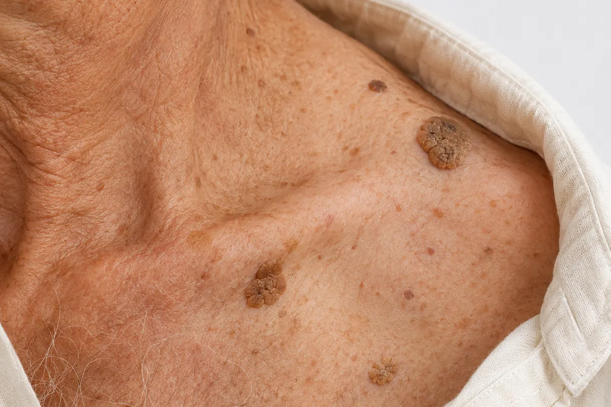
Seborrhoeic keratosis is one of the most common benign skin lesions after the age of 40. It looks like a patch or a small raised plaque, with a rough or waxy surface, which resembles a drop of wax “stuck” to the skin, and may be beige, brown or almost black. Although sometimes called “seborrhoeic wart” or “age wart”, it is not caused by a virus or contagious, and does not turn into cancer. The problem is another: some are dark and irregular and may look like a melanoma. On this page we explain how it is recognised, why it appears and why it tends to multiply, how it is told apart from other lesions, which methods exist to remove it when it bothers you and what to expect from each, and, prominently, when a lesion needs to be assessed before any decision. At Clínica Luxmed, in Algés, the doctor examines the lesion and defines the path at the assessment consultation.

Reviewed by Dr Michelle Campagnoli, clinical director

  • 40+age at which they usually multiply
  • 1assessment, always first
  • Nocommitment
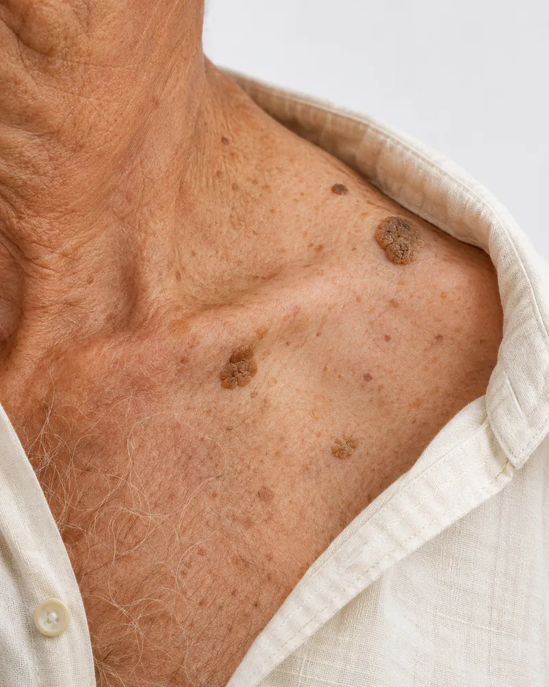
Collarbone area of a man in an open shirt, with seborrhoeic keratoses

How to recognise it

Is it a seborrhoeic keratosis?

These signs help to describe what you see and to prepare the conversation with the doctor. Several skin lesions, including those that deserve more attention, look like a seborrhoeic keratosis, and that is why examination always comes before any decision.

Tick what you recognise0/7

Tick the signs you recognise in your case. This is not a diagnosis, it is only meant to give you an idea.

A lesion that is dark, asymmetrical, with irregular edges or several colours, that has changed, grows quickly, bleeds, forms a sore that does not heal or is different from all the others, needs a medical assessment before any treatment. The same applies if many lesions appear suddenly. If in doubt, call the SNS 24 line (808 24 24 24). 112 is only for an emergency, for example bleeding that does not stop.

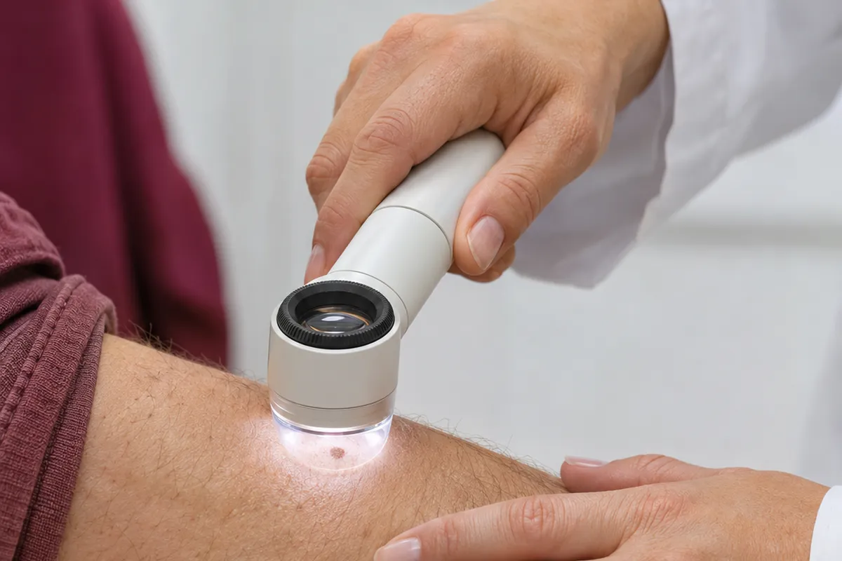
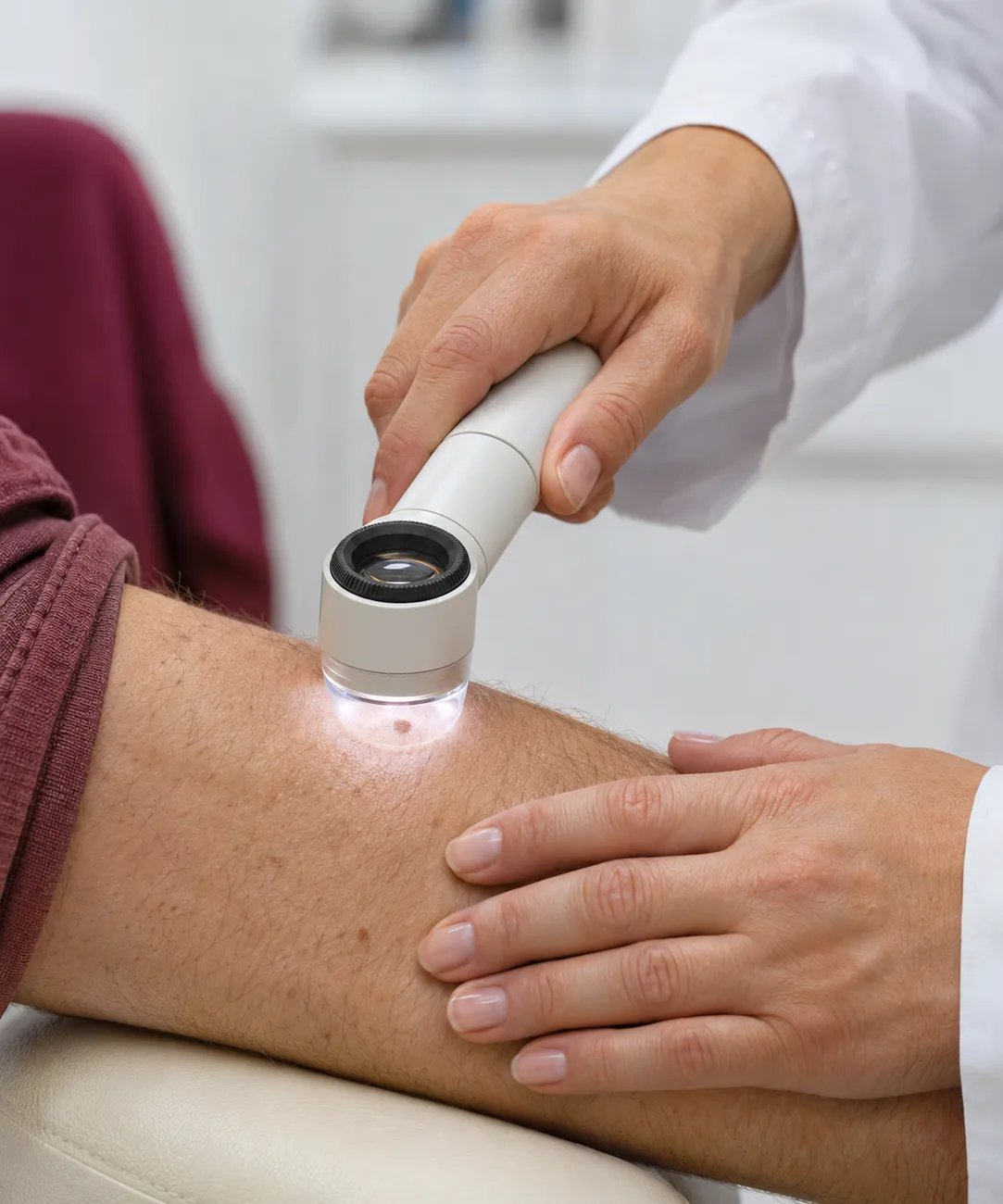
Female doctor examining a mole on the skin of the arm with a dermatoscope
Photograph of a consultation. The dots mark what the doctor is assessing.

Why it appears

What is behind seborrhoeic keratosis

The exact cause is not clear, but several factors are known that help to explain why some people have few and others have many. Choose one to see how it acts and what helps.

Age and skin ageing

Seborrhoeic keratosis is considered a degenerative change of the skin: it is rare before the age of 20, begins to appear around 30 or 40 and increases in number with the passing of time. That is why it is one of the most frequent lesions in people over 50, in men and women, and in all skin colours.

What favours it

The passing years: each decade usually brings more lesions, in general with the same appearance.

What helps

Knowing that it is a characteristic of mature, benign skin, and monitoring only those that change or stand out.

Normal or to be assessed

Does it look like a common keratosis or does it need assessment?

Most seborrhoeic keratoses are benign and not urgent, but there are situations in which nothing is removed or tried before a doctor has seen it. See which side your lesion is on.

  • What they look like. Plaques “stuck” to the skin, with sharp edges, beige to dark brown, with a rough or waxy surface, sometimes with small cysts or fissures, on the trunk, face, neck or scalp.

  • What stage they are at. They grow slowly, over months or years, and in general keep the same colour and appearance. They are not urgent and do not tend to change.

  • Why it is worth confirming. Some have an appearance similar to a melanoma, a basal cell carcinoma or an actinic keratosis, and only clinical examination, dermoscopy and, sometimes, analysis of the tissue tell them apart.

  • What usually helps. Leaving them be when they do not bother you, moisturising the skin, avoiding friction and, if they irritate or bother you aesthetically, booking an assessment to choose the method.

  • Tell us at the consultation. Since when they have been there, how many there are, whether any has changed, whether there are cases in the family, and which medication you take.

Not sure what it is? Book an assessment. The doctor examines the lesion, tells you what it is and whether it is advisable to monitor or remove it and, when there is doubt, refers to dermatology before any treatment.

Book an assessment

What can be done

Removing seborrhoeic keratosis: what to expect

Seborrhoeic keratosis does not need to be treated for health reasons. It is removed when it bothers you aesthetically, when it irritates with clothing or when there is doubt about the diagnosis, and the decision is always made after the lesion has been assessed. Among the methods described in medical guidelines are cryotherapy, for thin lesions, curettage and electrosurgery, for thicker lesions, laser, in selected cases, and localised chemical peel. All have drawbacks: there may be a lighter or darker patch, a scar, a need to repeat and, in a small trial on facial lesions, cryotherapy had lower satisfaction than electrosurgery and laser. Isolated lesions are removed easily, but there is no simple way of removing many at once, and others may appear with time. When the lesion raises doubts, removal is done in such a way that the tissue can be sent for analysis. At Luxmed, the path is defined at the assessment consultation, and the doctor refers to dermatology when there are doubts about the lesion.

Book an assessment
Dr Michelle Campagnoli, clinical director of Clínica Luxmed

First step

Assessment consultation

The doctor listens to what you are experiencing, examines your case and explains the suitable options. No treatment begins without this consultation.

you want to understand the cause and decide on the best path, with time and without commitment.

Book an assessment

What the journey looks like

From the first examination to follow-up

No pigmented lesion is removed without being assessed first. This is how it goes.

  1. 01At the clinic

    Discuss the history of the injury

    The doctor asks since when it has been there, whether it has changed, whether it bleeds or itches, how many there are and whether they appeared suddenly, whether there are cases in the family and which medication you take.

  2. 02At the clinic

    Examining and comparing

    She looks at the colour, surface and edges of each lesion and compares them with each other. If any stands out or raises doubt, the decision is different: referral to dermatology, dermoscopy and, if needed, analysis of the tissue.

  3. 03At the clinic

    Deciding between monitoring and removing

    She explains what to expect from each path, including the method, healing, the risk of a patch or scar and the possibility of other lesions appearing, and defines with you what makes sense.

  4. 04Over time

    Follow-up

    She agrees how to monitor the lesions that remain, how to photograph them and when to return, and sees the area again if any changes or if new lesions appear.

Day to day

What helps at home

What you do between consultations counts towards monitoring the skin well and not worsening a lesion that is not urgent.

The golden rule

First, knowing what it is

Most mistakes with keratoses begin with treating at home a lesion that nobody has confirmed. A benign keratosis is not urgent, but a lesion that looks like a keratosis and is something else is. If it changes, bleeds or stands out from the others, it is not for trying things on, it is for showing.

Looking at and photographing the lesions

A dated photograph makes it possible to compare colour, shape and size after a few months, and to see whether any has changed.

In practice: photograph in good light with a ruler or coin beside it, every two or three months, and bring the images to the consultation.

Protect the skin from the sun

The relationship with the sun is not proven, but sun protection reduces damage to the skin and makes it easier to detect lesions that change.

In practice: sunscreen every day on exposed areas, clothing and shade in the hours of most sun.

Avoiding friction on the lesions

An irritated keratosis becomes red, crusted and itchy, and may be misinterpreted.

In practice: clothing and straps that do not rub on the lesion, moisturising the skin and assessment of those that become irritated often.

Telling the doctor what changes

New lesions, a rapid increase in number or a lesion that stands out are a reason for a consultation, not for waiting.

In practice: bring the list of medicines, say how long ago the lesions appeared and which have changed.

Dr Michelle Campagnoli, clinical director of Clínica Luxmed

Who will look after you

An assessment carried out by the doctor

At Luxmed, the first step is always an assessment with Dr Michelle Campagnoli, at the clinic in Algés. She is the one who examines the lesions, confirms what they are and tells you whether it is advisable to monitor or remove, before any decision.

  • Physical examination and medical historyExamination of the lesions, their location and progress, the clinical history, the medication and any signs that call for another assessment, with referral to dermatology when there is doubt.
  • A plan tailored to youA clear plan, with the method to consider explained, the risks of patch or scar, the possibility of new lesions appearing and follow-up over time.
  • No obligationThe cost of any treatment is confirmed only after the assessment.
Book an assessment

Book a consultation

Book your consultation

Choose how you prefer to get in touch. We will arrange the assessment consultation with Dr Michelle Campagnoli.

Booking through the websiteAssessment consultationTalk to our assistant on WhatsApp and ask for the current price of the assessment.

New Life Miraflores, Algés · Office Center, R. Santa Teresa do Menino Jesus n.º 6, 9.º C, 1495-048

Your details are used only for this contact. Any treatment depends on a prior medical assessment.

Request an appointmentIt takes less than a minute.
Speciality
You do not need to go into detail. The doctor will talk to you during the consultation.
Request received. Our team will contact you as soon as possible.
What is seborrhoeic keratosis?

It is a benign skin lesion, beige, brown or almost black, with a rough or waxy surface and a “stuck-on” look, which appears with age, above all from the age of 40, on the trunk, face, neck and scalp. It is also called “seborrhoeic wart” or “age wart”, but it is not caused by a virus, is not contagious and does not turn into cancer. It increases in number with time and has a family component. Only examination confirms the diagnosis.

Can seborrhoeic keratosis turn into cancer?

No. It is a benign growth and is not considered a pre-malignant lesion. What may happen is that a skin cancer arises by chance beside or within a keratosis, and it is also possible to confuse a dark keratosis with a melanoma or another cancer. So, what matters is not that the keratosis turns into cancer, but telling one from the other: a lesion that has changed, bleeds or stands out should be assessed.

Is it contagious? Is it a wart?

It is not contagious and it is not a wart in the usual sense. The name “seborrhoeic wart” is misleading: warts are caused by the HPV virus, and seborrhoeic keratosis is not, does not pass from one person to another and does not spread by contact. Nor does it come from poor hygiene or oil in the skin. It is a benign change in the cells of the superficial layer of the skin.

Why do I have so many? Why do more keep appearing?

Because age and genetics are the main factors: the number increases with the years, and there are families in which having many is common. The sun and friction may play a role, but it is not proven. Having many is normal and does not mean that anything is wrong. What deserves attention is the sudden appearance of many lesions in a short time, or a lesion different from the rest, which call for assessment.

Seborrhoeic keratosis or melanoma: how can I tell them apart?

A keratosis is usually “stuck” to the skin, with a rough or waxy surface, a uniform colour, sometimes with cysts and fissures under the dermatoscope, and progresses slowly. A melanoma tends to be asymmetrical, with irregular edges, several colours, and may change quickly. The distinction is not always easy, even for doctors without dermoscopy. So you should not try to tell them apart on your own: if the lesion is dark, irregular or has changed, ask for assessment; if in doubt, a biopsy or removal with analysis is done.

When should I see a doctor about a keratosis?

When the lesion is very dark or irregular, grows quickly, changes colour or shape, bleeds, forms a sore that does not heal, is different from the others, or when many appear suddenly. Also before removing any pigmented lesion, and if a keratosis becomes irritated often. Call the SNS 24 line, 808 24 24 24. 112 is only for an emergency, for example bleeding that does not stop.

Do I have to remove seborrhoeic keratosis?

Not for health reasons. It is benign and many people leave them be. It is removed when it bothers you aesthetically, when it irritates with clothing or accessories, or when there is doubt about the diagnosis. If you are going to have it removed for aesthetic reasons, the decision is yours, after confirming what it is, and you should know the risks of each method. There is no hurry, and a stable lesion may simply be monitored.

Which methods exist for removing seborrhoeic keratosis?

Among the methods described in medical guidelines are cryotherapy (liquid nitrogen), for thin lesions; curettage and electrosurgery, for thicker lesions, with curettage allowing the tissue to be analysed; laser, in selected cases; and localised chemical peel. All have drawbacks: a lighter patch, sometimes permanent, above all in dark skin, a scar and a need to repeat. The same lesion rarely returns after being removed, but others may appear with time. At Luxmed, the path is defined at the assessment consultation.

Can I remove seborrhoeic keratosis at home? And creams?

You should not scrape, burn, cut or apply acids or corrosive products: there is a risk of infection, scarring, incomplete removal and, above all, of treating a lesion that turns out to be malignant. Moisturising creams soften the lesion, but do not remove it. There is a topical 40% hydrogen peroxide formulation assessed in trials, whose use should be decided by a doctor. Before any attempt, ask for assessment.

What should I bring and say at the assessment?

Say since when the lesions have been there, whether any has changed, bleeds or itches, how many there are and whether they appeared suddenly, whether there are cases in the family, which conditions you have and which medication you take. Bring old photographs, if you have them, for the doctor to see the progress. If you have a lesion that stands out, point it out straight away. Book the assessment in Algés, with no commitment.

We could not find that question. Ask us on WhatsApp

Scientific references

This page is based on the sources below and was reviewed by Dr. Michelle Campagnoli. Last updated on .

  1. Hafner C, Vogt T. Seborrheic keratosis. J Dtsch Dermatol Ges, 2008.
  2. Wollina U. Recent advances in managing and understanding seborrheic keratosis. F1000Res, 2019.
  3. Zaresharifi S, Robati RM, Dadkhahfar S, Forouzanfar M, et al. Efficacy and safety of cryotherapy, electrodesiccation, CO2 laser, and Er:YAG laser in the treatment of seborrheic keratosis. Dermatol Ther, 2021.
  4. Abbasi NR, Shaw HM, Rigel DS, Friedman RJ, McCarthy WH, Osman I, et al. Early diagnosis of cutaneous melanoma: revisiting the ABCD criteria. JAMA, 2004.
  5. Grob JJ, Bonerandi JJ. The 'ugly duckling' sign: identification of the common characteristics of nevi in an individual as a basis for melanoma screening. Arch Dermatol, 1998.
  6. American Academy of Dermatology. Seborrheic keratoses: overview. (aad.org, acedido 2026-11-11)
  7. American Academy of Dermatology. Seborrheic keratoses: diagnosis and treatment. (aad.org, acedido 2026-11-11)
  8. DermNet (Oakley A). Seborrhoeic keratosis. (dermnetnz.org, acedido 2026-11-11)
  9. American Academy of Dermatology. How to spot melanoma: the ABCDEs. (aad.org, acedido 2026-11-11)
  10. NHS. Symptoms of melanoma skin cancer. (nhs.uk, acedido 2026-11-11)
CallAssistantAvailableBook