Algés, Oeiras Mon to Fri · 09:00 to 13:00 and 14:00 to 19:00 · Sat · 09:00 to 14:00
WhatsApp Book a consultation

Body aesthetics

Moles on the skinAlmost all moles are benign, but only an examination confirms it, and only then is it decided whether it is worth monitoring or removing

Moles are small patches or pigmented dots that almost everyone has, and the great majority are benign. They appear mainly in childhood and adolescence, and new lesions, almost always harmless, continue to appear throughout life. What matters is knowing how to tell the common mole from one that deserves attention: the ABCDE rule, the “ugly duckling” mole and change over time help with this. On this page we explain how each situation is recognised, what may be behind a new mole, when it is an aesthetic decision and when it is a medical one, what to expect from a removal and, prominently, when a mole needs to be assessed before anything else. At Clínica Luxmed, in Algés, the doctor examines the moles and defines the path at the assessment consultation.

Reviewed by Dr Michelle Campagnoli, clinical director

  • ABCDEthe rule for monitoring moles on the skin
  • 1assessment, always first
  • Nocommitment
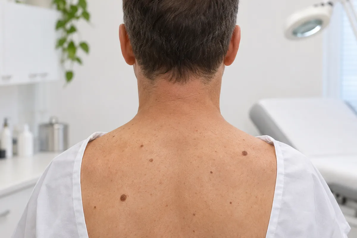
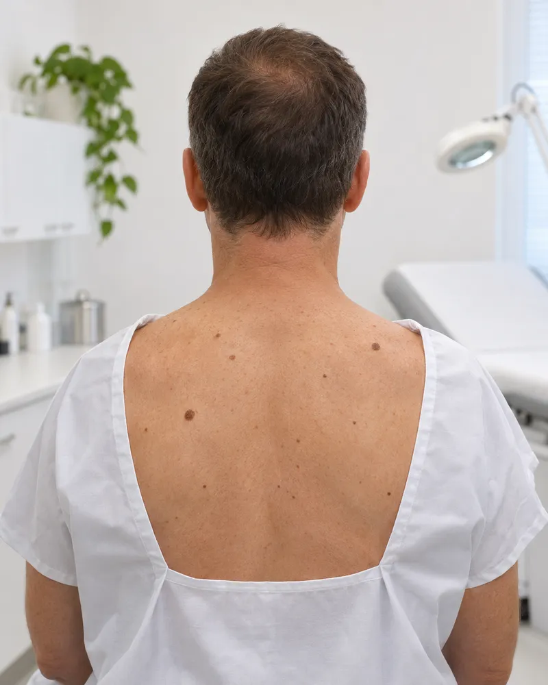
Back of a man in a consulting room, with brown moles

How to recognise it

Is this mole normal or does it deserve attention?

These signs help to describe what you see and to prepare the conversation with the doctor. They do not replace examination, which is the only way to know whether a mole is benign.

Tick what you recognise0/7

Tick the signs you recognise in your case. This is not a diagnosis, it is only meant to give you an idea.

A new mole in an adult, asymmetrical, with an irregular outline, several colours, that changes, itches, forms a crust or bleeds, or that is different from all the others, needs a medical assessment before any aesthetic procedure. Do not wait to see if it goes away. If in doubt, call the SNS 24 line (808 24 24 24).

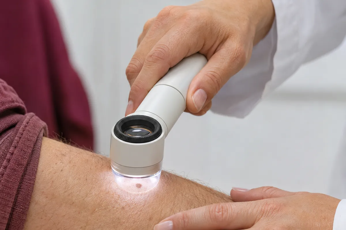
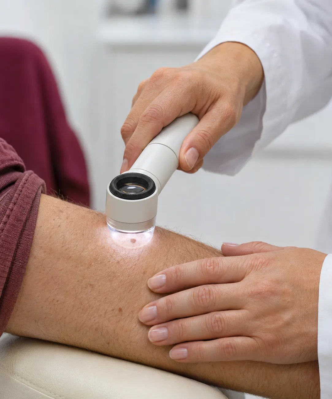
Female doctor examining a mole on the skin of the arm with a dermatoscope
Photograph of a consultation. The dots mark what the doctor is assessing.

Why they appear

What is behind moles on the skin

Moles result from skin cells that produce pigment and cluster together. There are factors that decide how many appear and which deserve more attention. Choose one to see how it acts and what helps.

The cells that give colour to the skin

A common mole, or naevus, is a small cluster of cells that produce pigment. It may be flat or raised, smooth or rough, and lighter or darker, and it is normal for an adult to have several. Some are present from birth, most appear in childhood and adolescence, and some fade with age.

What weighs on it

Having many moles or atypical moles, for example large, with an irregular outline or uneven colour.

What helps

Knowing your own moles and what is usual for each, so as to notice when something changes.

Normal or to be assessed

Does it look like a common mole or does it need assessment?

Most moles are benign and not urgent, but there are characteristics in which nothing is done or tried before a doctor has seen it. See which side yours is on.

  • What it looks like. Round or oval, with a smooth outline and uniform colour, flat or raised, and often similar to the other moles of the same person.

  • What stage it is at. Stable over time. It is normal for new moles to appear in childhood and adolescence, and for some to fade with age.

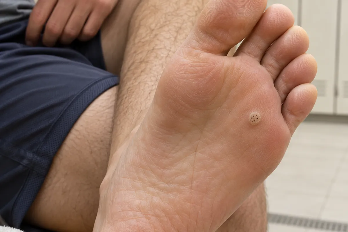
  • Benign age-related lesions. Seborrhoeic keratoses (brown, rough and with a “stuck-on” look), cherry angiomas (bright red dots), sun lentigines (flat brown patches) and soft fibromas (little skin tags) are benign and very common.

  • What usually helps. Examining the skin regularly, protecting yourself from the sun and, if a mole bothers you aesthetically or by rubbing, booking an assessment to decide.

  • Tell us at the consultation. Since when it has been there, whether it has changed, whether there is melanoma in the family, whether you have had sunburn or used sunbeds.

Not sure what it is? Book an assessment. The doctor examines the mole, tells you whether it is to be monitored, whether it is advisable to refer to dermatology or whether it may be removed for an aesthetic reason, and only then is the method decided.

Book an assessment

What can be done

Removing a mole: when it is aesthetic and when it is medical

The decision to remove a mole has two possible reasons. The aesthetic one, when the mole is benign and bothers by its look or by rubbing. The medical one, when the mole is suspicious, changes or bleeds. In both cases the rule is the same: assess first and, if the mole is removed, analyse the tissue. Among the methods described are excision with stitches, which allows the whole mole to be analysed and leaves a linear scar, shave removal of benign raised moles, which leaves a small scar and may recur, and laser or electrosurgery for benign non-pigmented lesions, which do not allow the tissue to be analysed. Methods that destroy the mole should not be used on pigmented moles without a diagnosis. Local anaesthesia is generally used, a small scar remains and the result of the analysis may take a few weeks. Removing a mole does not make it malignant. At Luxmed, the indication and the method are defined at the assessment consultation, with referral to dermatology whenever necessary.

Book an assessment
Dr Michelle Campagnoli, clinical director of Clínica Luxmed

First step

Assessment consultation

The doctor listens to what you are experiencing, examines your case and explains the suitable options. No treatment begins without this consultation.

you want to understand the cause and decide on the best path, with time and without commitment.

Book an assessment

What the journey looks like

From the first examination to follow-up

No mole is removed without being assessed first. This is how it goes.

  1. 01At the clinic

    Telling the story of the mole

    The doctor asks since when it has been there, whether it has changed, whether it itches or bleeds, whether there is melanoma in the family, how the skin reacts to the sun and whether you have had sunburn or used sunbeds.

  2. 02At the clinic

    Examining the moles

    She looks at the mole that worries you and at the others, looks for the characteristics of the ABCDE rule and the “ugly duckling”, and checks whether it is a mole or another benign age-related lesion.

  3. 03At the clinic

    Deciding between monitoring, removing or referring

    She explains what to expect from each path: monitoring and seeing again, removing for an aesthetic reason with analysis of the tissue, or referring to dermatology when there are suspicious characteristics.

  4. 04Over time

    Following up and monitoring

    She agrees with you how the moles are to be monitored, examines the skin again when needed and reviews the result of the analysis and care of the scar, if there was removal.

Day to day

What helps at home

What you do between consultations counts towards detecting early what has changed and protecting the skin.

The golden rule

Has it changed or is it new? Show it

Most mistakes with moles begin with waiting to see or with trying to remove at home a mole that nobody has assessed. A new mole, one that is different from the others or that has changed, itches or bleeds is not for trying things on or for waiting: it is for showing to a doctor.

Examining the skin regularly

Knowing your own moles is what allows you to notice when one changes or when a different one appears.

In practice: once a month, in good light and with a mirror, look at the whole body, including the back, scalp, palms of the hands, soles of the feet and nails.

Photographing the mole that worries you

A dated photograph helps to see whether the mole has changed, and the doctor can compare it at the consultation.

In practice: photograph it close up, with a ruler or coin beside it and good light, and bring the photographs to the consultation.

Protecting yourself from the sun

Accumulated sun and sunburn increase the risk of skin lesions.

In practice: shade between 11 and 15 h, clothing, a hat, sunglasses and sunscreen (minimum SPF 30), reapplied after swimming.

Telling the doctor your history

Melanoma in the family, fair skin, many moles and sunburn change how often the skin should be examined.

In practice: bring the information to the consultation and agree with the doctor how often you should come back.

Dr Michelle Campagnoli, clinical director of Clínica Luxmed

Who will look after you

An assessment carried out by the doctor

At Luxmed, the first step is always an assessment with Dr Michelle Campagnoli, at the clinic in Algés. She is the one who examines the moles, sees whether there are suspicious characteristics and tells you whether it is advisable to monitor, remove for an aesthetic reason or refer to dermatology, before any decision.

  • Physical examination and medical historyExamination of the moles that worry you and of the rest, their progress, the clinical and family history and sun exposure, before any procedure.
  • A plan tailored to youA clear plan, without promises: monitor, remove with analysis of the tissue or refer, with the risks and the expected scar explained, and follow-up over time.
  • No obligationThe cost of any treatment is confirmed only after the assessment.
Book an assessment

Book a consultation

Book your consultation

Choose how you prefer to get in touch. We will arrange the assessment consultation with Dr Michelle Campagnoli.

Booking through the websiteAssessment consultationTalk to our assistant on WhatsApp and ask for the current price of the assessment.

New Life Miraflores, Algés · Office Center, R. Santa Teresa do Menino Jesus n.º 6, 9.º C, 1495-048

Your details are used only for this contact. Any treatment depends on a prior medical assessment.

Request an appointmentIt takes less than a minute.
Speciality
You do not need to go into detail. The doctor will talk to you during the consultation.
Request received. Our team will contact you as soon as possible.
How do I know whether a mole on the skin is a concern?

The ABCDE rule helps: A for asymmetry (one half different from the other), B for irregular or poorly defined borders, C for several colours in the same mole, D for a diameter greater than a pencil eraser, although they may be smaller, and E for evolution, that is, a change in size, shape, colour or relief, with itching, crust or bleeding. An “ugly duckling” mole, different from all the others, and a new mole in an adult also deserve assessment. It does not replace medical examination: if in doubt, ask for assessment.

What is the “ugly duckling” mole?

It is the mole that does not look like the rest on the same body. Most people have moles with a similar appearance to one another, and the one that stands out for being larger, darker, lighter or of a different shape deserves to be examined. It is a complement to the ABCDE rule, which helps above all people with many moles.

Are moles that appear after 40 normal?

They are often benign age-related lesions: seborrhoeic keratoses (brown, rough and with a “stuck-on” look), cherry angiomas (red dots), sun lentigines (flat brown patches) and soft fibromas (little skin tags). But a new pigmented mole in an adult should be examined, because melanoma may also appear this way, and a rough lesion that does not go away with time may be an actinic keratosis, which calls for assessment.

Are red moles dangerous?

Cherry angiomas, small bright red dots, are benign and very common. But a red lesion that grows, bleeds, forms a crust or does not heal should not be presumed benign and should be seen by a doctor.

Is a mole with hairs or that itches safer or more dangerous?

Having hairs is not a reliable criterion: what matters is the ABCDE rule and change. Persistent itching or bleeding without a clear cause are warning signs and justify assessment, even if the mole looks normal.

Can I have a mole removed just for aesthetic reasons?

You can, if it is benign and after it has been assessed. It is a frequent decision when the mole rubs on clothing or bothers by its look. Before removing, the mole should be examined and, if removed, the tissue should be analysed. At Luxmed, the indication and the method are defined at the assessment consultation, with referral to dermatology whenever there are suspicious characteristics.

What methods exist for removing a mole?

Described are excision with stitches, which allows the whole mole to be analysed and leaves a linear scar, shave removal of benign raised moles, which leaves a small scar and may recur, and laser or electrosurgery for benign non-pigmented lesions, which do not allow the tissue to be analysed. Methods that destroy the mole should not be used on pigmented moles without a diagnosis. The choice depends on the mole, the area and the assessment.

Does removing a mole make it become malignant? Does it leave a scar?

No: it is a common myth that removing a mole makes it malignant. A small scar always remains, whose type depends on the method and the area. It is done with local anaesthesia, and the result of the analysis may take a few weeks. If the result calls for another assessment, the doctor explains the next steps.

When should I see a doctor about a mole?

Faced with any characteristic of the ABCDE rule, an “ugly duckling” mole, a new mole in an adult, or a mole that changes, itches, forms a crust or bleeds, and always before removing any mole. Call the SNS 24 line, 808 24 24 24. 112 is only for an emergency, for example bleeding that does not stop.

What should I bring and say at the assessment?

Say since when each mole that worries you has been there, whether it has changed, whether it itches or bleeds, whether there is melanoma in the family, whether you have fair skin, whether you have had frequent sunburn or used sunbeds. Bring old photographs, if you have them, for the doctor to see the progress, and show all the moles that worry you, even the less obvious ones. Do not apply products or try to remove anything beforehand. Book the assessment in Algés, with no commitment.

We could not find that question. Ask us on WhatsApp

Scientific references

This page is based on the sources below and was reviewed by Dr. Michelle Campagnoli. Last updated on .

  1. Abbasi NR, Shaw HM, Rigel DS, Friedman RJ, McCarthy WH, Osman I, Kopf AW, Polsky D. Early diagnosis of cutaneous melanoma: revisiting the ABCD criteria. JAMA, 2004.
  2. Grob JJ, Bonerandi JJ. The 'ugly duckling' sign: identification of the common characteristics of nevi in an individual as a basis for melanoma screening. Arch Dermatol, 1998.
  3. American Academy of Dermatology. What to look for: ABCDEs of melanoma. (aad.org, acedido 2026-11-11)
  4. NHS. Moles. (nhs.uk, acedido 2026-11-11)
  5. Green AC, Williams GM, Logan V, Strutton GM. Reduced melanoma after regular sunscreen use: randomized trial follow-up. J Clin Oncol, 2011.
  6. de Berker D, McGregor JM, Mohd Mustapa MF, Exton LS, Hughes BR. British Association of Dermatologists' guidelines for the care of patients with actinic keratosis 2017. Br J Dermatol, 2017.
  7. NHS. Actinic keratoses (solar keratoses). (nhs.uk, acedido 2026-11-11)
  8. American Academy of Dermatology. Seborrheic keratoses: overview. (aad.org, acedido 2026-11-11)
CallAssistantAvailableBook